














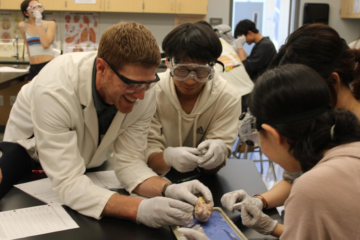
In Kristofer Orre’s fourth-period Anatomy and Physiology class, students stepped away from note-taking for a week to perform dissections on sheep brains and eyes from March 16-20.
Having previously done a sheep heart and pig fetus dissection prior to the brain and eye dissections, students found the brain and eye dissections much simpler, but just as valuable learning.
“The realness that comes from doing a dissection really affirms what is learned in those previous steps and helps students have a deeper understanding and appreciation for those structures,” Orre said.
The parts used for dissections are byproducts of the food industry, he said, meaning the brains and eyes are preserved and later sold to schools for educational use. When students dissect these structures, there is no blood, but there is a noticeable smell from the preservative.
For the eye dissection, students worked in pairs to remove fatty tissue and muscle remnants before using scissors to cut the eyes in half. By revealing the inside, students were able to see structures like the lens and pupil.
“I didn’t know the eye would be hollow in the inside, and I didn’t know [the vitreous humor] was like jelly around the lens, so that was really off-putting,” senior Mahika Jandhyala said. “But it was really valuable to see that eyes are built of so many complex structures. Perceiving one fraction of light takes so much work. It really helped going layer by layer to see what all the light passes through.”
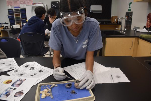
By dissecting the lens, students gained a better understanding of both the anatomy and physiology of the eye’s structure. For example, students can see sheep eye’s tapetum lucidum — the reflective, iridescent layer behind the retina.
The tapetum lucidum is what gives the sheep enhanced night vision. While a human’s eye does not contain this structure, students can draw parallels on how structure relates to function.
“Everybody takes away something different,” Orre said. “It’s this interesting mix between how delicate the structures are, but also how tough and resilient they are.”
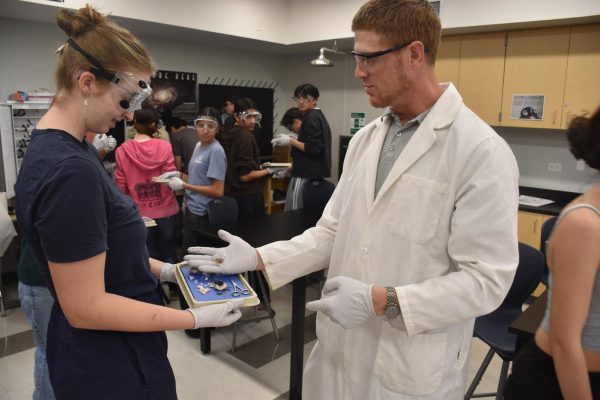
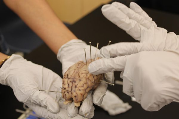
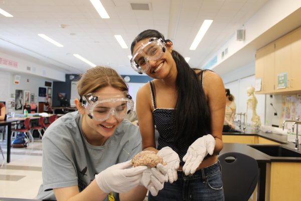
To start the sheep brain dissection, students teamed up in groups of four and removed the meninges, a shiny membrane protecting the brain. Then, they cut the brain in half to examine the left and right hemispheres.
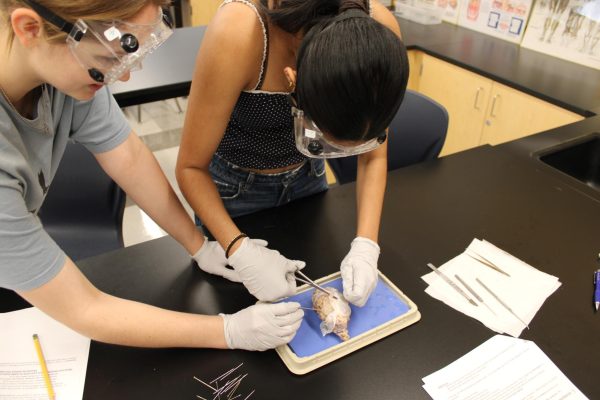
Students also tested their understanding by placing pins into specific regions of the brain. As they worked, Orre moved from table to table, checking each pin’s placement and guiding students on identifying structures.
(Grace Lin)
“Our models have these very clear boundaries that make it easy to see where one region ends and the next one begins,” Orre said. “It is those boundaries that are not easily discernible in the dissection, so you have to use a little bit of imagination and fall back on that prior experience of seeing it in models and pictures.”
Ultimately, the dissections in Orre’s class are part of a hands-on approach to learning anatomy. It allows students to connect structure to function and truly grasp the physiology part of the class.
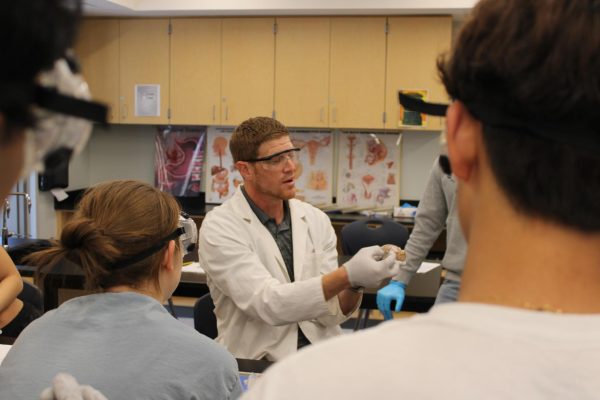
“You can learn a lot from a book, lectures and online resources, but nothing replaces holding it in your hand and seeing it for yourself,” Orre said.
