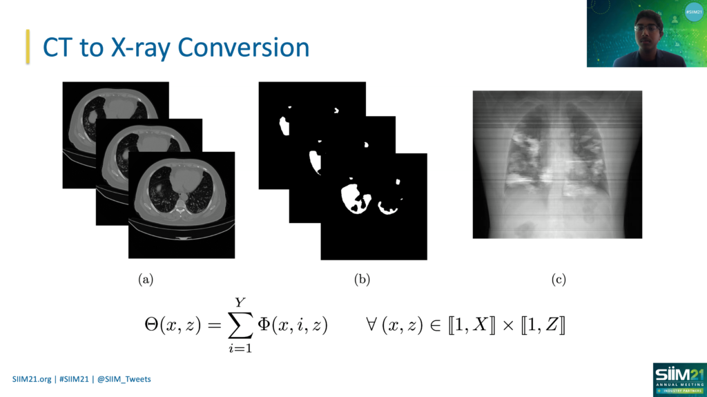
After senior Vignav Ramesh presented his research, “COVID-19 Lung Lesion Segmentation Using a Sparsely Supervised Mask R-CNN on Chest X-rays Automatically Computed from Volumetric CTs,” at the Society for Imaging Informatics in Medicine 2021 (SIIM21) Annual Meeting in May, he prepared for the questioning round from scientists attending the Zoom conference.
To his surprise, many asked Vignav about how his research could be applied in their own projects — a recognition of its utility in multiple areas of medical artificial intelligence (AI).
As a result of his work, Vignav received the 2021 Helen and Paul Chang Foundation New Investigator Travel Award during that conference. He became the first high schooler to ever receive the award in a pool of graduate students and PhD students competitors.
His winning research was guided by Stanford University Biomedical Informatics Professor Dr. Daniel Rubin and focused on measuring the severity of COVID-19 through the segmentation of X-rays on patients’ chests.
The project involved research in artificial intelligence and the medical field, two of Vignav’s passions.
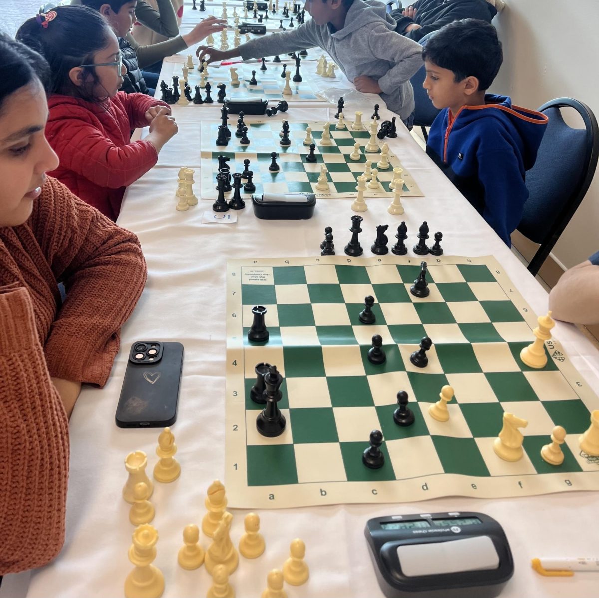
“I got into artificial intelligence through gaming because I thought it was really cool to use this technology to create automatic solutions to games like checkers, chess and more,” he said.
He learned the basics of AI through Stanford University’s Machine Learning course on Coursera. He then deepened his knowledge through participating in multiple hackathons and programming contests, creating medical-related projects involving artificial intelligence.
His older sister, Saratoga High Class of 2016 alumna Ashvita Ramesh, is now a medical student studying at the Feinberg Medical School in Northwestern University. She showed Vignav the projects she created in medical school and got Vignav interested in how AI can be used to facilitate medical work.
“I noticed that a lot of things that she was doing could be streamlined with AI and deep learning,” Vignav said. “I reached out to a couple of different professors at various universities, and Dr. Rubin responded to me. That’s how I started working in his lab.”
Vignav and Rubin developed the idea for this project upon realizing that, although computed tomography (CT) scans are effective data for COVID-19 severity quantification, they are not affordable for most patients. Instead, they found that such quantification could be achieved through X-rays, which are more cost effective and conducive to lung lesion identification. To do this, they used segmentation, a process of outlining regions of interest in the scans using edge-detection AI.
Though the research is only in academia at the moment, the publishing of this research will allow more doctors to look into the possibility of using X-Rays and segmentation. Vignav and his team are looking to put the research into use at hospitals, but implementation of new technologies requires multiple rounds of testing and improvements.
“I think the intersection of medical imaging and AI is a really important topic of research that I’m really interested in,” Vignav said. “Much farther into the future, I hope to see this model integrated clinically, revolutionizing healthcare in the U.S. and abroad.”


